Figure 2.
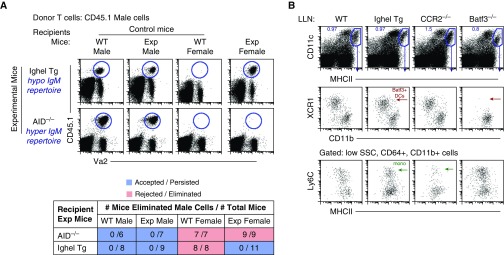
Natural IgM antibodies are essential for the initial recognition and elimination of neoantigen-expressing cells. (A) Flow data illustrate three controls: two negative controls (acceptance, male cells into male WT and KO/transgenic [Tg] mice) and one positive control (rejection, male cells into WT female mice). Exp mice are IghelMD4 (hypo-IgM repertoire) AID−/− (hyper-IgM repertoire) female mice. Data represent two independent experiments consisting of n = 3–5 animals per group. Table displays combined experiments with the number of mice that rejected male cells (numerator) over the number of total mice examined (denominator). Acceptance is shown in blue, rejection in red. (B) Lung-draining LN (LLN) flow plots. Top row: live cells plotted as CD11c versus MHCII to illustrate migratory DCs, gated blue. Middle row: migratory DCs are plotted as XCR1 versus CD11b to illustrate the two overarching DC subtypes: XCR1+, CD11blow Batf3+ DCs (red arrow) and CD11b+, Irf4+ DCs. Batf3−/− mice lack Batf3+ DCs, whereas IghelMD4 (Ighel) mice contain Batf3+ DCs. Bottom row: LN cells pregated on low side scatter (SSC), CD64+, CD11b+ cells and plotted as Ly6C versus MHCII to illustrate LN Ly6C+ monocytes. CCR2−/− mice display significantly diminished Ly6C+ monocytes (green arrow), whereas IghelMD4 mice display a normal quantity of LN monocytes.

