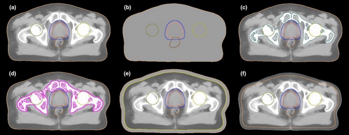
Figure 2.

Illustration of the original sCT and the sCTs with errors introduced. (a) original sCT, (b) all tissue assigned as water, (c) bone structure assigned as cortical bone, (d) enlarged bone structure with 2 mm cortical bone, (e) enlarged patient body contour with +10 mm water and (f) decreased patient body contour with −10 mm. The delineations in images are PTV (blue), CTV (pink), femoral head (yellow) and rectum (brown).
