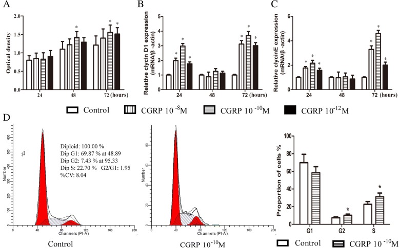
Fig. 1.
CGRP stimulated the proliferation of EPCs in dose- and time-dependent manners. After 24 h, 48 h, and 72 h of incubation with or without a series of concentrations CGRP (10− 8–10− 12 M) in high-glucose DMEM with 10% FBS, cell viability was assessed by the optical density via CCK-8 staining (a). Q-PCR was used to detect two proliferation-related genes, cyclin D1 (b) and cyclin E (c). Flow cytometry analysis of the cell cycle after 72 h of incubation (d). Data represent the means ± SEM of three experiments. * p < 0.05 compared with control

