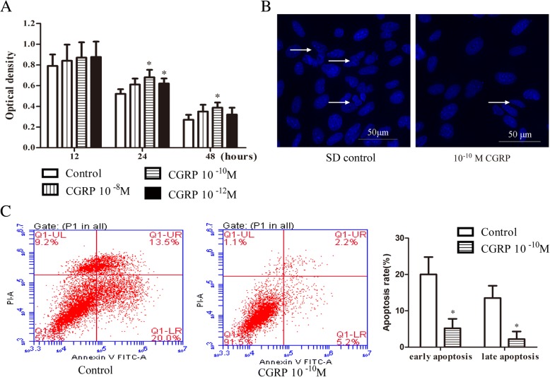
Fig. 2.
CGRP inhibited EPC apoptosis. After 12 h, 24 h, and 48 h of incubation with or without CGRP (10− 8–10− 12 M) in high-glucose DMEM during SD-induced apoptosis, cell viability was assessed by the optical density and by CCK-8 staining (a). The protective effects of 10− 10 M CGRP on the morphology of cell nuclei were evaluated by DAPI staining; the arrow indicates a cell nucleus that has undergone condensation and fragmentation (b). Cell apoptosis of control EPCs and EPCs stimulated with 10− 10 M CGRP after 48 h was analysed by flow cytometry analysis (c). Data represent the means ± SEM of three experiments. * p < 0.05 compared with control

