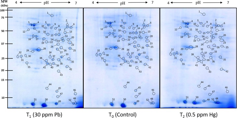
Fig. 6.
Representative 2D proteome maps of soybean root nodules of control, Pb-treated (30 ppm) and Hg-treated (0.5 ppm). 400 μg of total protein was loaded on to 11 cm/non-linear/pH 4–7 IPG strips for first dimensional run on IEF cell followed by second dimensional run on 12% SDS-PAGE. The proteins were visualised by staining of gels with blue silver stain. The encircled protein spots with numbering represents differentially expressed proteins which were considered for tryptic digestion and subsequent identification through MALDI-TOF MS/MS

