. 2018 Jul 26;9(37):7338–7353. doi: 10.1039/c8sc02470g

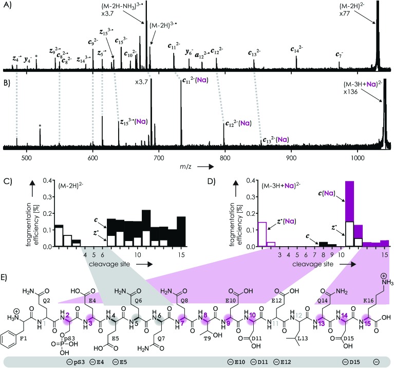
Fig. 1. Spectra from niECD of (A) (M – 2H)2– and (B) (M – 3H + Na)2– ions using a skimmer potential of 10 V (asterisks indicate harmonic signals)70 and (C, D) corresponding site-specific fragmentation efficiency (N–Cα backbone bond cleavage, >98% c, z˙ and <2% c˙, z fragments); (E) primary structure of the phosphopeptide with proposed protonation sites (N terminus and K16) and possible locations of the four negative charges (pS3, E4, E5, E10, D11, E12, D15, and the C terminus) indicated below; highlighted in gray are sites from which no fragments were observed in niECD of (M – 2H)2– ions, and in purple sites at which fragment ion formation was substantially reduced or prevented in niECD of (M – 3H + Na)2– ions.

Fig. 1