Figure 4.
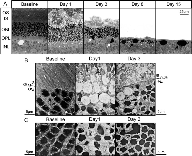
Light-induced alterations in photoreceptor nuclei and ONL. Plastic sections were obtained from retinas of I307N Rho mice before and 1, 3, 8, or 15 days after exposure to 20,000 lux of light for 30 minutes. (A) Representative visual fields with an original magnification of 40× are displayed. Unlike the samples from the day 3 and day 8 time points that contained subretinal detachments, the RPE was adherent to the ONL in the day 15 sample. The RPE was cropped out of images for simplicity. White arrows: large pleomorphic nuclear material. Black arrows: large spherical nuclear material. (B, C) Representative visual fields are displayed with an original magnification of 5000× of the: (B) interface between the ONL and subretinal space, and the (C) ONL. (B) Obliteration of the OLM was apparent by day 1 after light exposure. The ONL was separated from the inner segments by nuclei-free spaces surrounded by membrane or ECM. Deposition of dense ECM in the ONL occurred by day 3 and contributed to a newly formed demarcation between the ONL and subretinal space. (C) The normal configuration of chromatin at baseline was replaced by condensed chromatin consistent with apoptosis. The tight packing of the photoreceptor nuclei was disrupted, especially on day 1.

