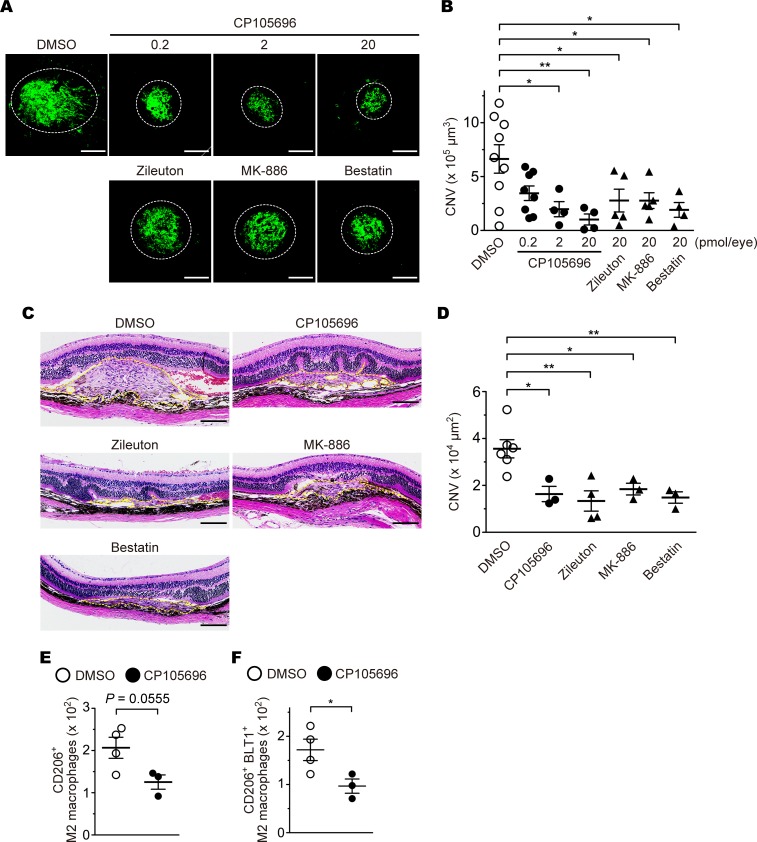
Figure 9. The effects of a BLT1 antagonist and LTB4 synthesis inhibitors on development of CNV.
Images of iB4 staining (A) and CNV volume (B) in the RPE-choroid from aged WT mice (>20 weeks old) after administration of 0.2–20 pmol of CP105696 (a BLT1 antagonist; filled circles) or 20 pmol of zileuton (a 5-LO inhibitor), MK-886 (a FLAP inhibitor), or bestatin (a LTA4H inhibitor) (filled triangles), or vehicle (open circles). n = 4–9 mice per group. (C and D) Images of H&E staining (C) and CNV lesion area (D). H&E staining of the retinas after aged WT mice (>20 weeks old) with laser-induced injury were treated with CP105696, zileuton, MK-886, and bestatin (20 pmol/eye) or DMSO. Yellow dotted lines show the lesion area. n = 3-6 per group. (E and F) The number of the ocular-infiltrating CD206+ or CD206+BLT1+ M2 macrophages was analyzed on day 3 after laser injury from CP105696- (20 pmol/eye) or DMSO-injected WT mice (> 20 weeks old). n = 3–4 per group. Scale bar: 100 μm (A and C). *P < 0.05; **P < 0.01 (1-way ANOVA with Dunnett’s post hoc test [B and D] and Student’s t test [E and F]). Results are representative of at least 2 independent experiments (A–D).

