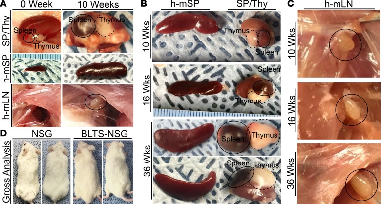
Figure 2. Lymphoid tissues development in the BLTS humanized mouse model.
(A) Representative gross analysis of the human spleen and thymus organoids (SP/Thy; images not presented at similar scale) in the kidney capsule, humanized murine spleen (h-mSP; images presented at similar scale), humanized murine lymph nodes (axillary) (h-mLN; images presented at similar scale) in BLTS humanized mice at 0 weeks (day of transplantation) and 10 weeks after transplantation. Representative gross analysis of the (B) humanized murine spleen (h-mSP) and the human spleen and thymus organoids (SP/Thy) in the kidney capsule (images presented at similar scale) and (C) humanized murine lymph nodes (axillary) (h-mLN) in BLTS humanized mice at indicated time points after transplantation. (D) Representative gross analysis of the absence of graft versus host disease (lack of fur loss or wasting syndrome) in age-matched, nontransplanted NSG mice and BLTS humanized NSG mice at 36 weeks after transplantation. Shown are representative gross tissues or animals (n = 4 per group). Black circles identifies tissues of interest.

