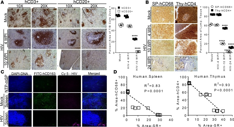
Figure 7. HIV-induced immunodeficiency correlates with lymphoid tissue fibrosis in the BLTS humanized mouse model.
(A and B) Human-specific immunohistochemical analysis of the kinetics of immunodeficiency in human lymphoid tissues, (A) T (Brown stain, hCD3+) and B (Brown stain, hCD20+) cell depletion in lymphoid follicle zones in human spleen organoid and (B) macrophages (Brown stain, CD68+) and CD4+ T (Brown stain, CD4+) cell depletion in human spleen (SP) and thymus (Thy) organoids, respectively, following inoculation at 1 × 10⁵ IU per mouse in BLTS humanized mice; mock inoculated mice, age-matched to the 24 weeks post-infection (24 WPI) served as controls. Immunohistochemical staining of spleens from nontransplanted NSG mice with the human antibodies were nonreactive (data not shown). (C) In situ hybridization (RNA expression) analysis of macrophage (CD163+, FITC, green fluorescence) levels and the presence of HIV viral RNA (red/pink fluorescence) in the human spleen organoid in mock-inoculated and chronic HIV-infected (24 weeks after infection) BLTS humanized mice; spleen from nontransplanted NSG mice (NTP-mSP) served as control. (D) Correlation analysis of human macrophage or CD4+ T cell depletion and collagen deposition (Sirius red stained regions, % Area SR+) in human lymphoid tissues (human spleen and thymus organoids). Representative images are shown (n = 4 per group), and data is presented as mean values ± SEM. P values (****P < 0.0001) were determined using 1-way ANOVA between more than 2 groups for each immune cell staining or lymphoid tissue, with mock as the control group. R2 coefficient was determined using linear regression and 16 mice. Scale bars: 200 μm.

