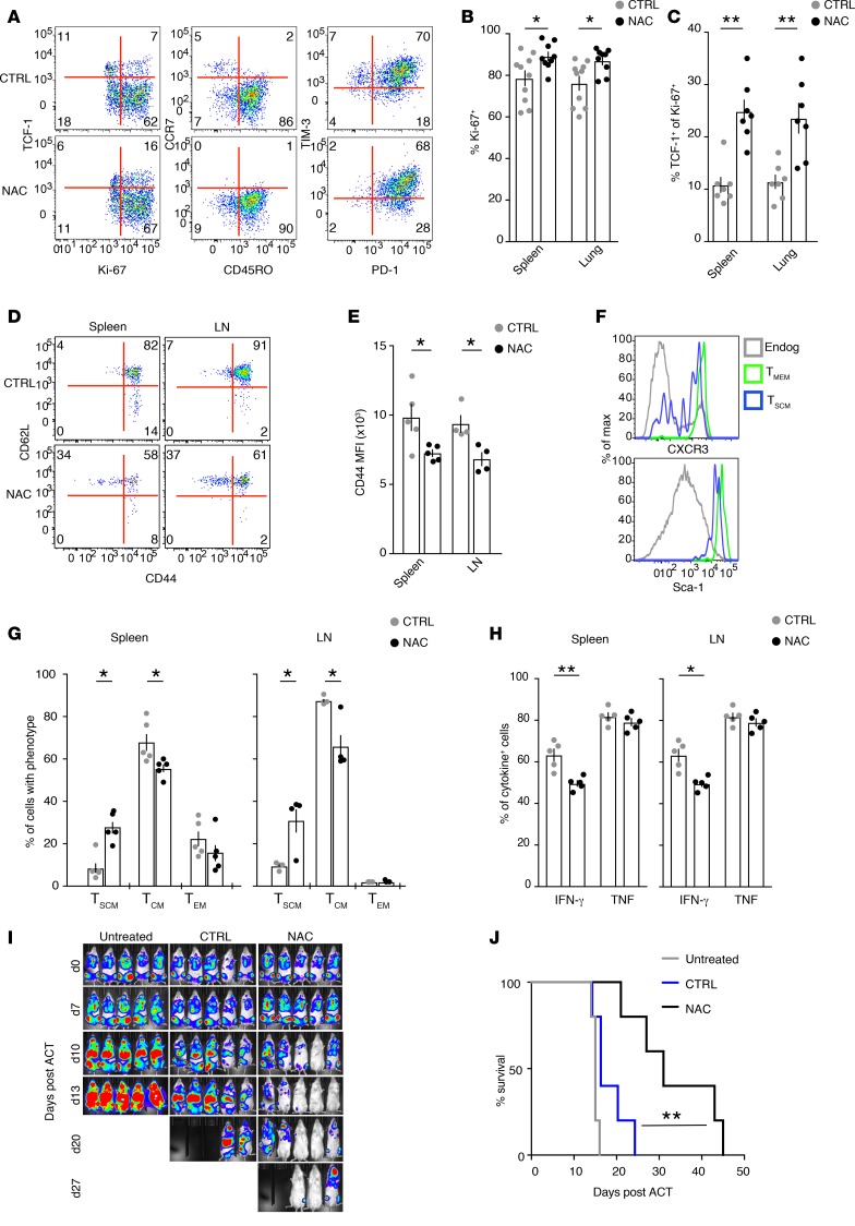
Figure 5. NAC promotes stem-like memory formation and potent antitumor immunity in vivo.
(A) Representative FACS analysis of protein expression by human circulating CD8+ Tn cells, activated as in Figure 2A, and adoptively transferred in NSG mice. Data refer to CD8+ T cells isolated from spleens at day 15 after ACT. Similar data were obtained from n = 2 additional exp. (n = 3–4 mice/group). (B and C) The ex vivo frequencies of Ki-67+CD8+ T cells (B; CTRL, n = 10; NAC, n = 9; n = 3 exp.) and TCF-1+ cells within the Ki-67+CD8+ T cell fraction (C; CTRL and NAC, n = 7; n = 2 exp.), as obtained in spleens and lungs of mice treated as in A. (D) Representative CD44 and CD62L expression in HBV Cor93-specific CD8+ Tn cells activated in vitro with cognate peptide and high-dose IL-2 for 8 days followed by transfer in wild-type C57/BL6 mice (n = 5 mice/condition in n = 1 exp.). Data refer to day 35 after ACT. Transferred cells were identified by the congenic marker CD45.1 while endogenous cells were CD45.2. (E) MFI (mean ± SEM) of CD44 in CD8+ T cells from D. (F) Representative histograms of surface CXCR3 and Sca-1 expression by Cor93-specific CD8+ T cells gated as Tscm (CD44loCD62Lhi) or Tmem (CD44hiCD62Lhi) cells from an animal receiving NAC-treated CD8+ T cells. Endogenous host CD8+ T cells are shown as a control. Data refer to day 35 after ACT. Similar data were obtained from n = 4 more mice. (G) Frequencies (mean ± SEM) of Tscm, Tcm, and CD44hiCD62Llo Tem cells (gated as in D) within the Cor93-specific CD8+ T cells previously activated in the absence (CTRL) or presence of NAC. Data refer to day 35 after transfer (n = 5 mice/treatment group). (H) The frequency of Cor93-specific CD8+ T cells (adoptively transferred as in D) producing IFN-γ and TNF following restimulation with the cognate peptide for 4 hours (n = 5/group). (I) Bioluminescence imaging of NALM-6 leukemic cells in NSG mice left untreated or adoptively transferred with human CD8+ Tn cells previously activated with anti-CD3/28, IL-2, and IL-12 in the absence (CTRL) or presence of NAC for 8 days and redirected with CD19 CAR (n = 5 mice/group). (J) Kaplan-Meier survival curve of mice treated as in H. For all FACS dot plots, numbers indicate the percentage of cells identified by the gate. Statistical analyses were performed with nonparametric Mann-Whitney test (B, C, E [LN], and G), parametric Student’s t test (E [spleen] and H) or Mantel-Cox analysis (J). *P < 0.05; **P < 0.01.

