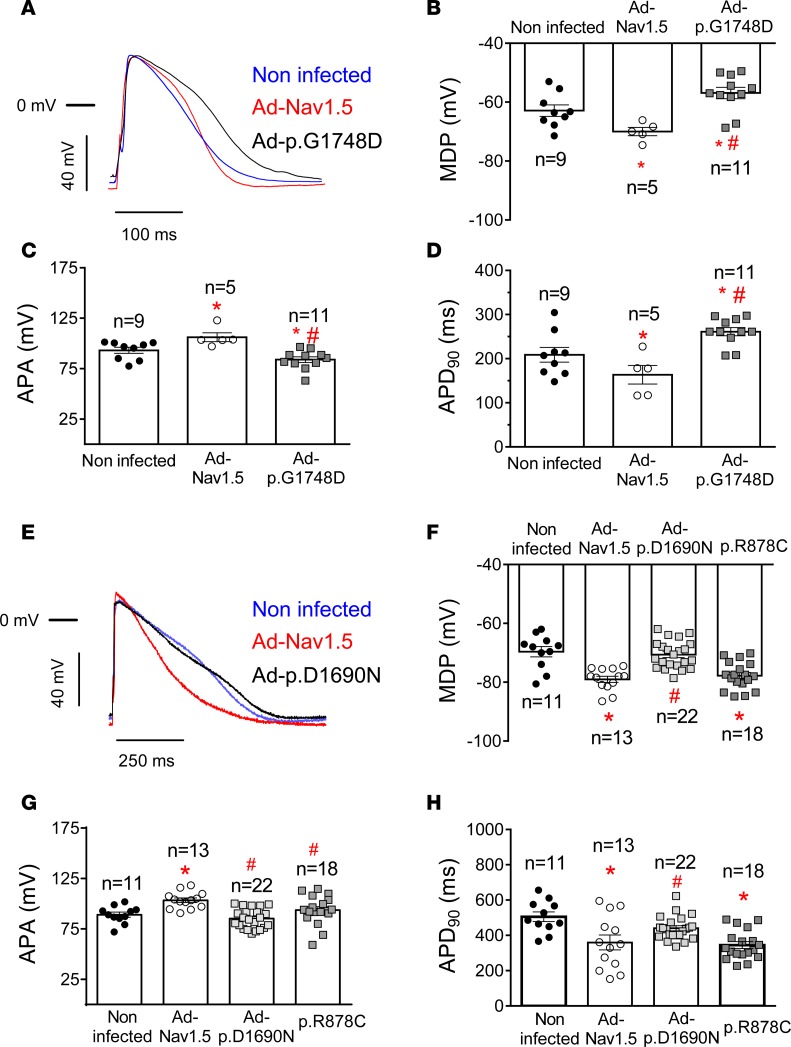
Figure 6. Effects of trafficking-defective Nav1.5 mutants on action potentials recorded in human induced pluripotent stem cell–derived cardiomyocytes.
(A) Superimposed action potential (AP) traces recorded in noninfected and Ad-Nav1.5– or Ad-p.G1748D–infected DF19-9-11T human induced pluripotent stem cell–derived cardiomyocytes (hiPSC-CMs) driven at 1 Hz. (B–D) Mean maximum diastolic potential (MDP) (B), AP amplitude (APA) (C), and AP duration measured at 90% (APD90) (D) for APs recorded at 1 Hz in noninfected hiPSC-CMs (black circles) and hiPSC-CMs infected with Ad-Nav1.5 (white circles) or Ad-p.G1748D (gray squares). (E) Superimposed AP traces recorded in noninfected and Ad-Nav1.5– or Ad-p.D1690N–infected iCell2 hiPSC-CMs driven at 1 Hz. (F–H) Mean MDP (F), APA (G), and APD90 (H) for APs recorded at 1 Hz in noninfected hiPSC-CMs (black circles) and hiPSC-CMs infected with Ad-Nav1.5 (white circles) or Ad-p.D1690N (light gray squares) or transfected with the cDNA encoding p.R878C Nav1.5 (dark gray squares). In B–D and F–H, each bar represents mean ± SEM of n experiments from at least 5 different dishes, and each dot represents 1 experiment. One-way ANOVA followed by Newman-Keuls and multilevel mixed-effects model were used for comparisons. *P < 0.05 vs. noninfected; #P < 0.05 vs. Ad-Nav1.5.

