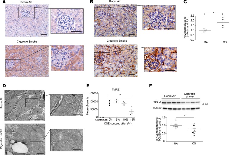
Figure 2. CS induces oxidative stress and mitochondrial injury in a murine model of COPD.
(A) Nitrotyrosine and (B) 8-Oxo-2′-deoxyguanosine (8-oxo-DG) staining detected by immunohistochemical staining of kidney tissue of mice exposed to room air (RA) or cigarette smoke (CS) for 6 months. Scale bars: 50 μm and 25 μm (magnified images). (C) Quantification of Nrf2 expression measured by Western blot in kidney tissue from mice exposed to RA or CS for 6 months (n = 4 in each group) normalized to β-actin and control. Data are mean ± SEM. *P < 0.05, analyzed by Student’s t test. (D) Transmission electron microscopy (TEM) of kidney sections harvested after 6 months of CS exposure, depicting mitochondrial swelling and loss of cristae definition, compared with RA control. Scale bars: 1 μm and 0.5 μm (magnified images). (E) Quantification of mitochondrial membrane potential of HK-2 cells treated in vitro with increasing concentrations of CS extract (CSE), measured by tetramethylrhodamine ethyl ester (TMRE) detected by FACS. Data are mean ± SEM. *P < 0.05 by 1-way ANOVA with Bonferroni’s post hoc test. (F) Representative Western blot of mitochondrial transcription factor A (TFAM) expression in kidneys after 6 months of CS exposure (3 independent determinations), with quantification (n = 8 per group). Data were normalized to TOM20 and RA control. All data are mean ± SEM. *P < 0.05 by 2-tailed Student’s t test.

