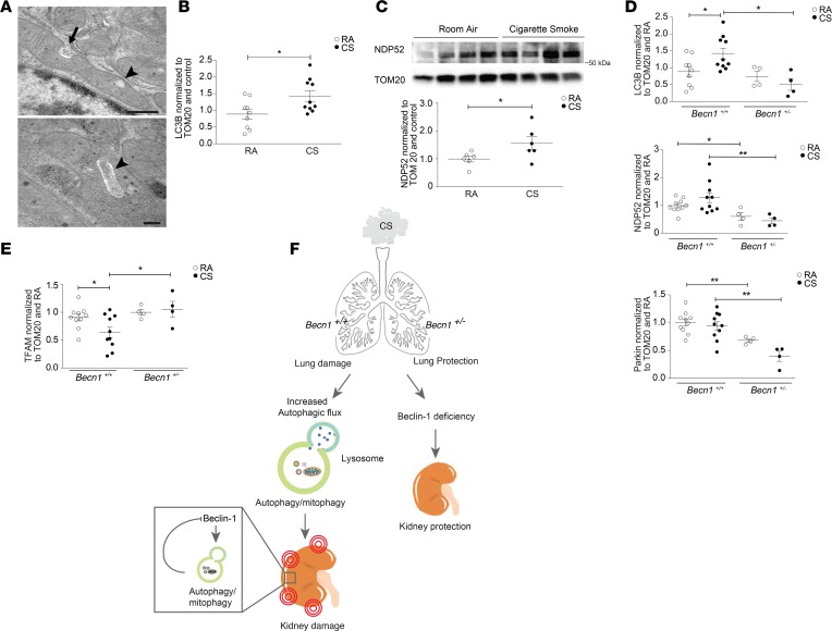
Figure 7. Becn1+/– mice have decreased mitophagy at baseline that does not increase after cigarette smoke exposure.
(A) Mitochondria of renal tubular epithelial cells of WT mice exposed to cigarette smoke (CS), showing invaginations (arrow) and loss of mitochondrial matrix (arrowhead) relative to RA control. Scale bars: 500 nm (top) and 200 nm (bottom). (B) Quantification of LC3B detected in mitochondrial fractions from WT mice, normalized to TOM20 and room air (RA) control (n = 9 for RA, n =10 for CS). (C) Representative Western blot of NDP52 in mitochondrial fractions from WT mice exposed to 6 months of CS compared with RA. Graph represents quantification of Western data normalized to TOM20 and RA control (n = 9 for RA, n = 10 for CS). (D) Quantification of Western blots of LC3B, NDP52, Parkin and (E) Mitochondrial transcription factor A (TFAM) expression detected in mitochondrial fractions after 6 months of CS or RA exposure in Becn1+/– mice relative to Becn1+/+ mice. Data were normalized to TOM20 and control (n = 10/group for Becn1+/+ mice, and n = 4/group for Becn1+/– mice). All data are mean ± SEM. *P < 0.05, **P < 0.01, analyzed by Student’s t test (B and C) and 1-way ANOVA with Bonferroni’s post hoc test (D and E). (F) Diagram representing CS-induced kidney injury. After systemic exposure to CS, autophagy is upregulated in kidneys in association with kidney injury. Becn1+/– mice have reduced autophagy and display reduced kidney injury after CS exposure. The increased autophagic activity and autophagosome turnover in response to CS exposure reduces Beclin-1 steady-state levels, as a potential compensatory mechanism.

