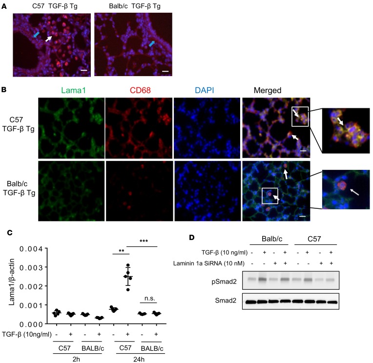
Figure 2. Strain- and cell-dependent Lama1 expression in lungs from TGF-β Tg mice.
Lungs from doxycycline-treated TGF-β1 Tg mice on the noted genetic backgrounds were evaluated. (A) Representative Lama1 IHC evaluation comparing C57 and BALB/c TGF-β Tg mice. White arrow indicates Lama1 positive macrophages while blue arrow indicates Lama1 stained area of basement membrane (lightly stained compared to macrophages) (B) Double IHC using anti-Lama1 (green) and anti-CD68 (red) antibodies. Arrows indicate cells stained with, Lama1, CD68 (macrophage specific marker), or both. Scale bars in A and B: 25 μm. (C) Fibroblasts were established from lungs of C57 and BALB/c mice and treated with TGF-β1 for 2 or 24 hours. The levels of Lama1 mRNA were evaluated by qRT-PCR. (D) Western blot evaluation on Smad2 activation in fibroblasts from C57 and BALB/c mice with and without TGF-β stimulation. A, B, and D represent evaluations in a minimum of 3 mice. Values in C represent mean ± SEM of evaluations in a minimum of 5 mice in each group. **P < 0.01, ***P < 0.001 compared with controls with no TGF-β treatment.

