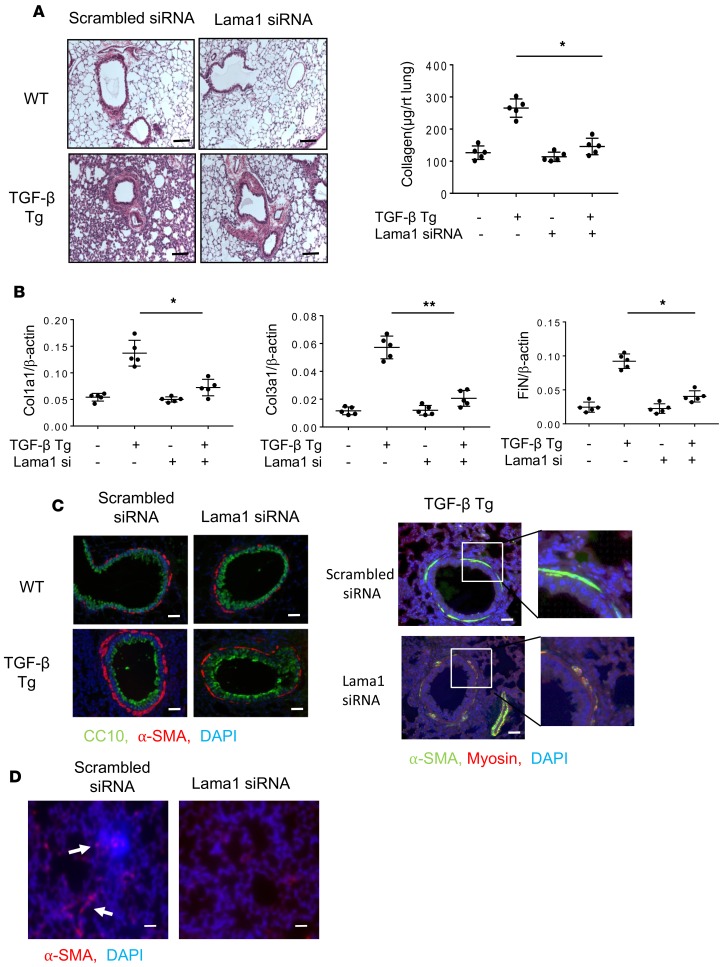
Figure 3. Lama1 plays an essential role in TGF-β–stimulated pulmonary fibrosis.
Lungs from 8-week-old WT and TGF-β1 Tg mice were evaluated after 2 weeks of Tg induction by doxycycline in drinking water. (A) Histologic evaluation with Mallory trichrome staining (left) and evaluation of lung collagen content with Sircol assays (right) comparing WT and Tg lungs from mice treated with control scrambled siRNA and Lama1 siRNA. (B) qRT-PCR evaluation of the levels of mRNA encoding extracellular matrix proteins (collagen type1 alpha1 [Col1a1], Col3a1, and fibronectin [FiN]) in lungs from WT and Tg mice treated with control scrambled siRNA or Lama1 siRNA. (C) Representative fluorescence double-label IHC evaluation of α–smooth muscle actin (α-SMA, red), CC10 (green), and nuclei (DAPI, blue) in lungs from WT and TGF-β1 Tg mice treated with scrambled control siRNA or Lama1 siRNA (left panels). Double-label IHC evaluation of α-SMA (green), myosin (red), and nuclei (DAPI, blue) in lungs from TGF-β1 Tg mice treated with scrambled siRNA control or Lama1 siRNA (right panels). (D) α-SMA IHC staining of the lungs of TGF-β1 Tg mice with scrambled siRNA or Lama1 siRNA silencing. Arrows indicate positively stained cells in the interstitial area in the lungs of C57 TGF-β1 Tg mice. A (left) and C are representative of evaluations in a minimum of 3 mice. The values in A and B represent mean ± SEM of evaluations in a minimum of 5 mice each group. *P < 0.05, **P < 0.01. Scale bars: 100 μm (A); 25 μm (C and D).

