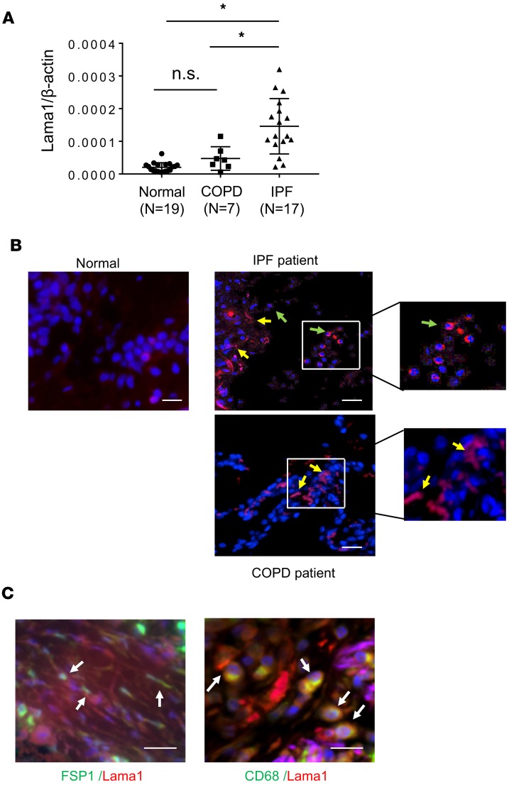
Figure 6. Increased expression of Lama1 in lungs from IPF patients.
(A) Levels of mRNA encoding Lama1 were evaluated, using qRT-PCR, in lung tissues from normal controls, patients with COPD, and patients with IPF. (B) Representative IHC staining of Lama1 in the lungs from controls and patients with IPF and COPD. Arrows indicate Lama1+ cells. Yellow arrows, fibroblasts-like cells; green arrows, macrophage-like cells. (C) Representative double IHC staining using macrophage- (CD68) and fibroblast-specific markers with Lama1 in lungs from IPF patients. *P < 0.05. Scale bars in B and C: 50 μm.

