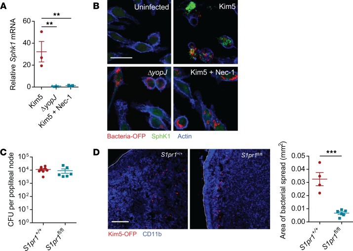
Figure 5. Necroptosis of infected macrophages alters local S1P gradients promoting intranodal bacterial spread.
(A) Fold change of Sphk1 mRNA in J774A.1 macrophages at 8 h.p.i. relative to uninfected controls (n = 3). (B) Immunofluorescence staining for SphK1 (green) in uninfected, Kim5-OFP–infected or in ΔyopJ-infected macrophages. Scale bar: 25 μm. (C) Bacterial numbers in PNs from mice whose mononuclear phagocytes were S1PR1-sufficient (Cx3cr1-Cre S1pr1+/+) or -deficient (Cx3cr1-Cre S1pr1fl/fl) 24 hours following footpad infection with Kim5 Y. pestis (n = 6). (D) Immunofluorescence staining of PNs of Cx3cr1-Cre S1pr1+/+ or Cx3cr1-Cre S1pr1fl/fl mice, 24 hours after footpad infection with Kim5-OFP bacteria. Scale bar: 50 μm. Graph shows area of bacterial spread measured from the PN images (n = 4–6). Data were analyzed via unpaired 2-tailed Student’s t test or 1-way ANOVA. Data are representative of at least 2 independent experiments. **P < 0.01, ***P < 0.001.

