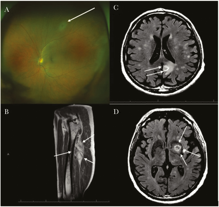
Figure 1.
A, Fundus photo of a left eye. Superior area of retinal whitening consistent with retinitis (white arrow) without overlying vitritis noted on exam. B, T1: magnetic resonance image (MRI) of the right thigh showing right thigh myositis (white arrows, increased fluid signal within the vastus intermedialis muscle). C and D, Brain T2 fluid-attenuated inversion recovery (FLAIR) MRI showing postcontrast multiple ring-enhancing lesions of toxoplasmosis (white arrows).

