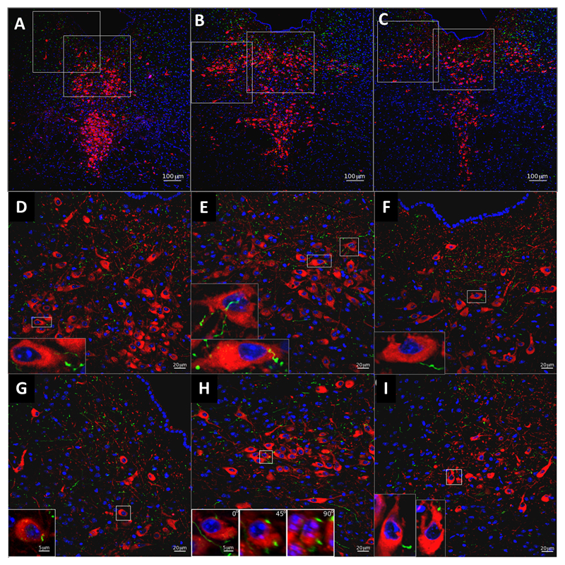
Fig 6.
Immunohistochemical labeling for YFP in YFP–PPG neurons (green) and 5-HT in serotonin neurons (red) in coronal sections through the DR nucleus of YFP–PPG mice. Low magnification micrographs showing the rostro-caudal extent of the DR examined (A-C). Groups of red 5-HT-immmunoreactive cell bodies are shown at higher magnification in D-I. Higher magnification of the dorsal subregion of the DR is displayed in D-E, and of the lateral subregion in G-I. For each of these subregions examples of individual TPH-positive neurons receiving YFP-labeled innervation from GLP-1-producing neurons are shown in the bottom left corner of each panel. Innervation of chosen individual neurons was confirmed by creating a three dimensional reconstruction of Z-stack images (Fig S3). High levels of immunoreactive YFP fibers are found in both the dorsolateral and dorsal section of the DR but not in the ventral section (Fig S4).

