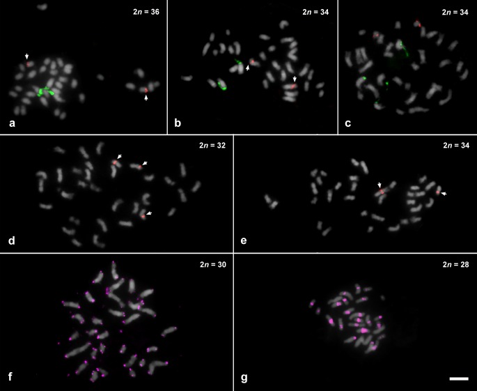
Fig 2. Distribution of repetitive sequences in species of Thaumatophyllum and Philodendron species, counterstained with DAPI and pseudocolored in gray (35S rDNA in green, 5S rDNA in red, and telomeric probe in pink).
(a) T. lundii; (b) T. mello-barretoanum; (c) P. angustilobum; (d) P. bipennifolium; (e) P. glaziovii; (f) P. giganteum; (g) P. callosum. Bar in g represents 5 μm.

