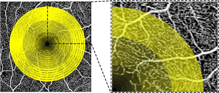
Fig 1. Multiple concentric rings grid used to analyze the choriocapillaris images in the study.
Optical coherence tomography en-face 3X3 mm angiogram of the superficial plexus with the superimposition of the grid used for the analysis of the choriocapillaris in this study. The superficial angiogram was used as a reference to manually center the grid on the center of the foveal avascular zone. The box on the right (inset of the image on the left), shows a magnified view of the superonasal quadrant of the grid. The concentric rings of the grid are defined by progressively wider concentric circles starting from the fovea and ending at a diameter of 2.5 mm (in the 3X3 scans) and 5 mm (in the 6X6 scans). Note, the spacing between circles decreases with greater distance from the fovea in order to maintain more similar areas between the various concentric rings.

