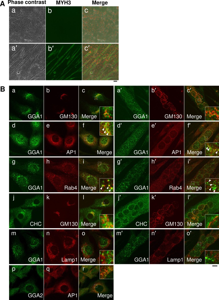
Fig 2. GGA1 is distributed from the Golgi to endosomes in C2C12 cells during myogenesis.
(A) Myogenic differentiation of C2C12 cultured myoblasts. C2C12 cells cultured in growth medium (day 0) and shifted to differentiation medium for four days (day 4) were stained with anti-myosin heavy chain 3 antibodies. (B) C2C12 cells cultured in the growth medium (day 0, a-o) and differentiation medium for four days (day 4, a′-o′) were subjected to immunofluorescent microscopy using anti-golgi associated, gamma adaptin ear containing, ARF binding protein 1 (GGA1) (a, c, d, f, g, i, m, o, a′, c′, d′, f′, g′, i′, m′, and o′), anti-golgin subfamily A member 2 (GM130) (b, k, b′, and k′), anti-adaptor related protein complex 1 subunit gamma 1 (AP1G1) (AP1: e, n, e′, and n′), anti-Ras-related protein rab-4A (RAB4) (h and h′), anti-clathrin heavy chain (CHC) (j and j′), and anti-lysosomal associated membrane protein 1 (LAMP1 (n and n′) antibodies. The cells from day 0 were stained with anti-GGA2 (p and r) and anti-AP1G1(q and r). All the images from anti-GGA1 or GGA2 antibodies are shown in green and those from other antibodies are shown in red. Merged images are indicated. Scale bar: 10 mm.

