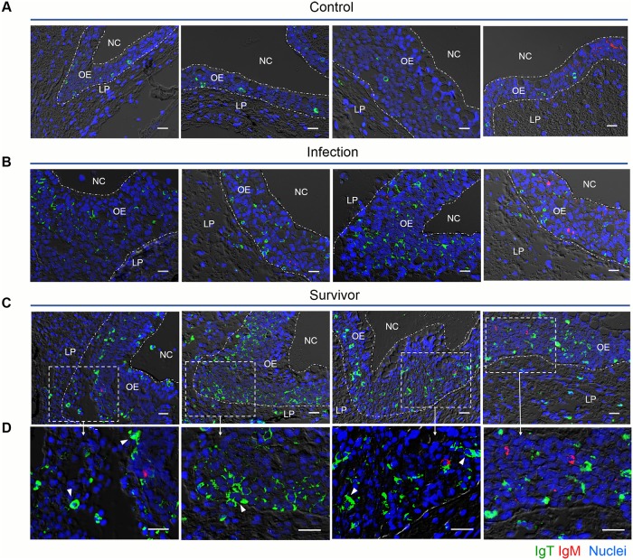
Fig 5. Accumulation of IgT+ B cells in the olfactory organ of trout infected with Ich.
DIC images of immunofluorescence staining on trout nasal paraffinic sections from uninfected fish (A), 28 days infected fish (B) and survivor fish (C), stained for IgT (green) and IgM (red); nuclei are stained with DAPI (blue). (D) Enlarged images of the areas outlined in c are showing some IgT+ B cells possibly secreting IgT (white arrowhead) (isotype-matched control antibody staining, S1B Fig). NC, nasal cavity; OE, olfactory epithelium; LP, lamina propria. Scale bar, 20 μm. Data are representative of at least three different independent experiments (n = 8 per group).

