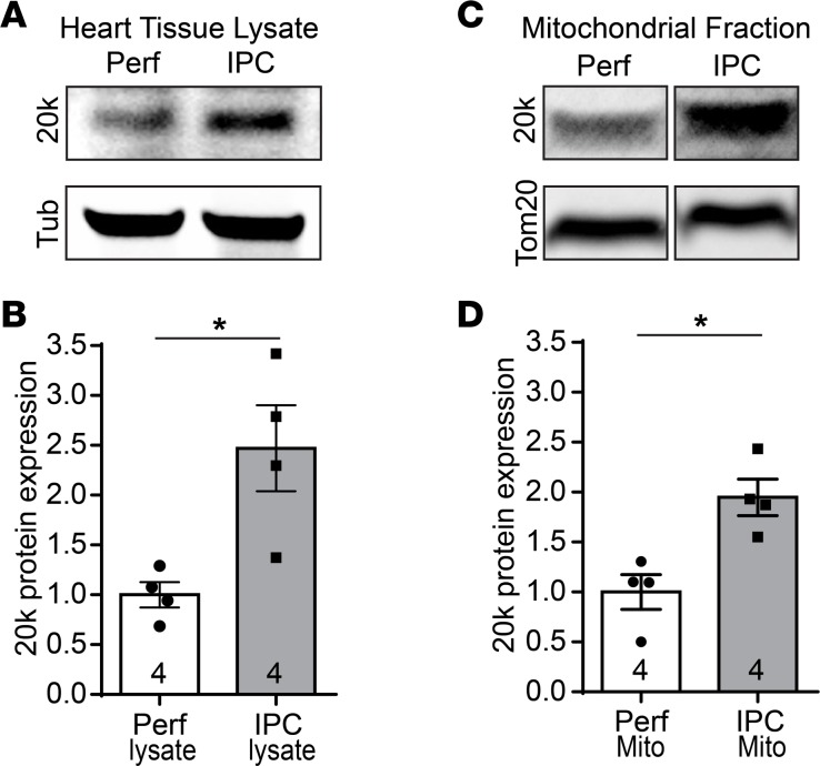
Figure 9. GJA1-20k increases with ischemic preconditioning.
Western blots of total tissue lysates (A) or F3 mitochondrial fractions (C) isolated from Langendorff-perfused mouse hearts, which were subjected to either ischemic preconditioning (IPC) or continuous perfusion (Perf) as a control. The blots are probed with a Cx43 C-terminus antibody to detect endogenous GJA1-20k, with a tubulin antibody, or with a Tom20 antibody. (B) Protein expression for GJA1-20k in A is normalized to tubulin and shown as fold change. n = 4 hearts per group. Data are shown as mean ± SEM. *P < 0.05, Mann-Whitney U test. (D) Protein expression for GJA1-20k in C is normalized to Tom20 and shown as fold change. n = 4 hearts per group. Data are shown as mean ± SEM. *P < 0.05, Mann-Whitney U test.

