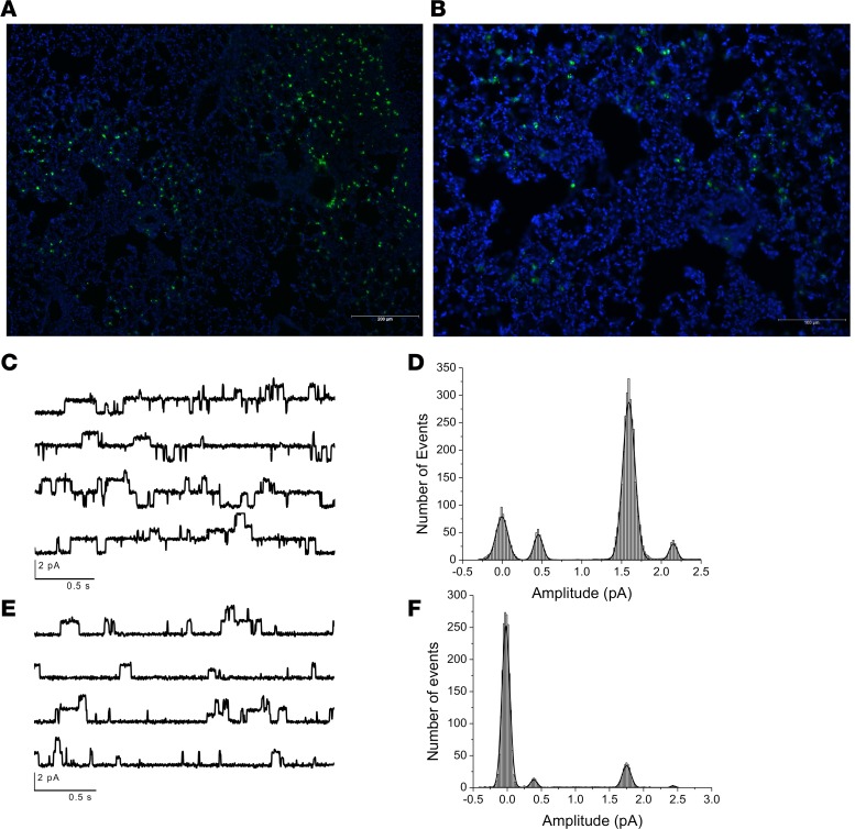
Figure 2. IAV reduces ENaC single-channel activity in infected but not in uninfected cells.
Mice were infected with 4,000 PFU of PR8ΔGFP, lungs were harvested 5 days p.i., and fresh 250 μM slices were prepared using a live tissue slicer from uninfected controls and infected animals as previously described (14). Slices were incubated in culture medium for 3–4 hours before ion channel activity was recorded from GFP+ and GFP- cells. Lung cryosections display widespread infection. GFP- cells exhibited single channels with conductance of 4 pS and 18 pS. Recordings were obtained at Vpatch + 100 mV when Vm was 0 mV (slices incubated in 135 mM KCl Ringer). Original magnification, ×100 (A) and ×200 (B). (C) Tracings of ENaC open probability conductance in GFP- cells. Histogram shown in D shows number of events for the 4 Ps channel only. (E) A representative recording of channel activities recorded from GFP+-infected AT2 cells. (F) GFP+ AT2 cells exhibited substantial reduction of 4 pS and 18 pS activity, without affecting their unitary conductances. Please see Figure 3 for quantification of these findings.

