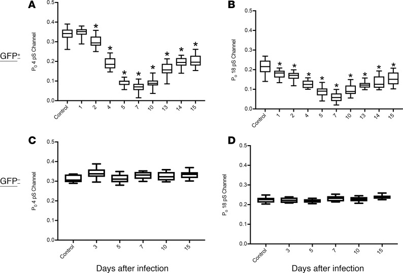
Figure 3. ENaC channel dysfunction during IAV infection.
Mice were infected with 4,000 PFU of PR8ΔGFP; lungs were harvested at days 1, 2, 4, 5, 7, 10, 13, 14, and 15 p.i.; and live slices were cut using a live tissue slicer. ENaC activities in infected AT2 cells (GFP+) were recorded using cell-attached mode of the patch-clamp technique. (A and B) Reduction of ENaC open probabilities for various times after infection. N = 5 mice and n = 29 cells per group. The maximum inhibition was seen at day 7, after which a slow recovery of both conductances was observed. (C and D) ENaC activity, measured as open probability (Po), was not affected in noninfected cells. Data are plotted as mean open probabilities, top and bottom quartiles ± SEM. Data were analyzed by 1-way ANOVA and post hoc Bonferroni correction for multiple comparisons. *P < 0.0001 for each time point compared with noninfected control values.

