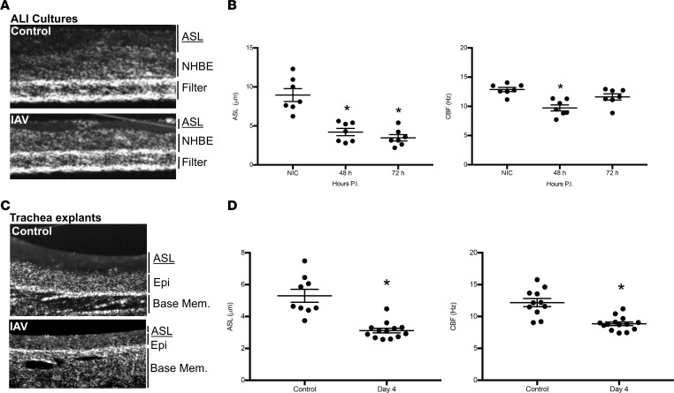
Figure 9. IAV infection reduces ASL depth and ciliary beat frequency in airway epithelial cultures and in vivo murine infection.
NHBEs were grown at an air-liquid interface and infected with A/California/07/2009 at a MOI of 1 for 48 or 72 hours. ASL depth and CBF were imaged using μOCT. (A) Representative OCT images from control or IAV-infected Transwell inserts identifying ASL, cell (NHBE), and filter structures. (B) IAV significantly (P < 0.003) reduced ASL depth and CBF within NHBE cultures at all time points and for the duration of experiments. (C) Representative images are displayed from μOCT imaging of tracheas. (D) ASL and CBF in tracheas from infected mice were significantly decreased after 4 days of infection. (B and D) Data, n = 7–13 per group, are plotted as mean ASL depth or CBF ± SEM. Data were analyzed by 1-way ANOVA and post hoc Tukey test for multiple comparisons. *P < 0.003 compared with noninfected controls.

