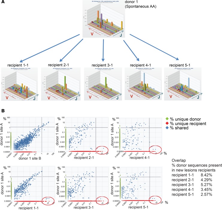
Figure 2. The majority of expanded T cell clones in recipient lesions are newly primed T cells.
Skin from a donor with spontaneous AA (donor 1) was grafted onto 5 recipient mice, which subsequently developed AA. The TCRβ repertoire of donor skin and new lesions in recipients was sequenced and plotted as a 3D histogram of variable versus joining region genes versus number of reads (counts) (A). The percentages of all detected TCRβ sequences in donor and recipient skin samples were plotted, showing sequences shared between donor and recipient (percentage shared [blue]), sequences unique to donor (percentage on y axis [green]), and sequences unique to recipient (percentage on x axis [red]). The most expanded clones from the donor skin are detected at lower frequencies in recipient lesions (top left quadrant), and the most expanded clones in the recipient lesions are primarily unique to the recipient (bottom right quadrant, red circle) (B). The percentage of donor sequences present in new recipient lesions is indicated (Overlap).

