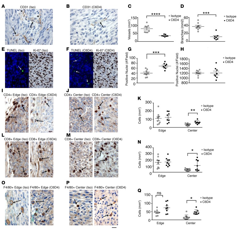
Figure 3. β8 expression increases tumor vessel growth, promotes tumor cell survival and immune exclusion.
(A–R) Established MC38 tumors were therapeutically treated with isotype (Iso) or C6D4 (10 mg/kg i.p. on days 0, 3, and 6) and harvested when tumors reached endpoint volume (2,000 mm3). Tumors were immunohistochemically assessed for vascular growth (A–D), apoptosis and proliferation (E–H), and immune infiltration (I–R). Representative 400× fields from tumor centers are shown for CD31+ vascular staining of (A) isotype-treated (Iso-treated) (A) or C6D4-treated (B) MC38 tumors. Arrows indicate representative CD31+ vessels, and in A, asterisks indicate representative branch points; (C) vascular density and (D) vessel branching assessed morphometrically from five 200× fields. Open boxes, isotype; filled boxes, C6D4. (E and F) Representative 400× fields from MC38 tumor centers from mice treated with isotype (E) or C6D4 (F) stained by TUNEL (green fluorescence, TUNEL+ (arrows) and blue, DAPI; left panels) or Ki-67 (right panels) and assessed for apoptosis (G) or Ki-67 positivity as a proliferation marker (H). (I, J, L, M, P, Q) Representative 400× fields from MC38 tumor edge (I, L, O) or center (J, M, P), as indicated above each photomicrograph, from mice treated with Iso (left panel) or C6D4 (right panel), as indicated above each figure, stained with anti-CD4 (I and J), anti-CD8 (L and M), or anti-F4/80 (O and P). Representative positively stained immune cells are indicated by arrows. Scale bar: 25 μm. Quantification from individual tumors for CD4+ T cells (K), CD8+ T cells (N), or F4/80 + macrophages (Q) from the tumor edge or center are shown. n = 8, in each antibody treatment group. Open boxes, isotype control; filled boxes, C6D4. Significance determined by unpaired Student’s t test. *P < 0.05, **P < 0.01, ***P < 0.001, ****P < 0.0001.

