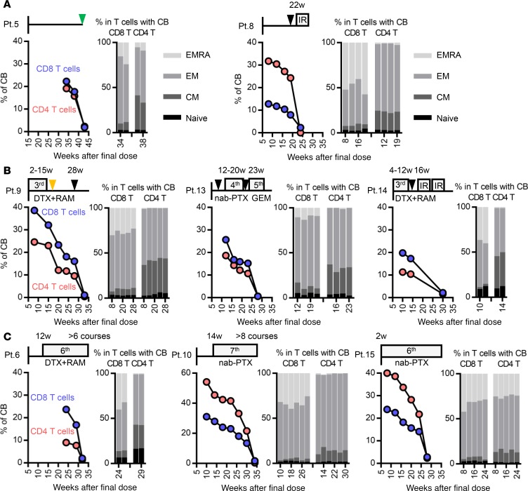
Figure 4. Following up binding status and differentiation markers in nivolumab-bound T cells in patients who underwent sequential treatment.
Fresh whole blood samples from 8 non–small cell lung cancer patients were followed up in terms of percentage of complete binding of nivolumab (left) and the ratio of 4 subsets (CD45RA+CCR7+ naive, CD45RA–CCR7+ central memory [CM], CD45RA–CCR7– effector memory [EM], and CD45RA+CCR7– effector memory RA [EMRA]) in the CB population (right) of CD8 and CD4 T cells. (A) Pt. 5 and 8 discontinued nivolumab treatment due to irAE. (B) Pt. 9, 13, and 14 and (C) Pt. 6, 10, and 15 discontinued nivolumab treatment due to progressive disease. (C) Pt. 6, 10, and 15 were well controlled by subsequent chemotherapies for more than 24 weeks. Black and green triangles indicate the points of progressive disease and tumor marker re-elevation without an increase in the size of the targeted tumor (as determined by CT scan), respectively. The orange triangle indicates the point of an adverse event of chemotherapy.

