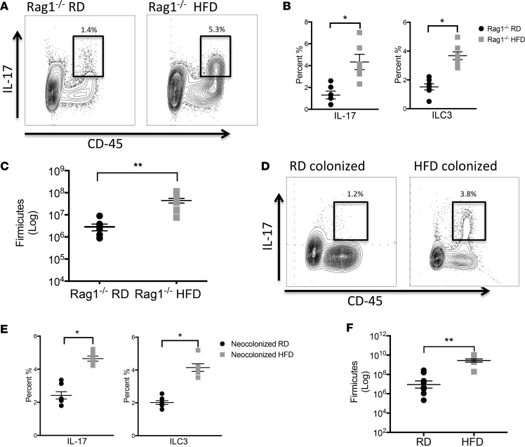
Figure 3. Rag1–/– mice on HFD have an expanded population of IL-17–producing ILC3s.
(A) Flow cytometric analysis of lamina propria (LP) cells from 2-week-old RD Rag1–/– and HFD Rag1–/– offspring was performed. Live cells were stained for CD45 (x axis) and IL-17 (y axis) and representative panels are shown here. Cells positive for CD45 and IL-17 are highlighted in the bold boxes. (B) Percentage of CD45+IL-17+ cells (left) and percentage of IL-17+Rorγt+CD4–CD127+NKp46+CD117+ ILC3s (right) in 2-week-old RD Rag1–/– and HFD Rag1–/– offspring. (C) Quantification of Firmicutes (log scale) in the small intestine of 2-week-old RD Rag1–/–and HFD Rag1–/– offspring by qRT-PCR. (D) Flow cytometric analysis of LP cells from 2-week-old neocolonized RD and neocolonized HFD offspring was performed. Cells positive for CD45 and IL-17 are highlighted in the bold boxes. (E) Percentage of IL-17+ cells and IL-17–producing ILC3s in 2-week-old neocolonized offspring. (F) Quantification of Firmicutes (log scale) in the small intestine of 2-week-old neocolonized RD and neocolonized HFD offspring by qRT-PCR. The data shown are representative of 3 experiments with 7–10 mice in each group, and are depicted as the mean ± SEM. *P < 0.05; **P < 0.01 by 2-tailed Student’s t test. HFD, high-fat diet; RD, regular diet.

