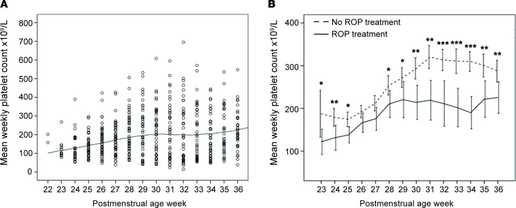
Figure 1. Platelet count and severity of retinopathy of prematurity.
(A) Platelet counts of all infants screened for retinopathy of prematurity (ROP) per postmenstrual age week (PMA) (each dot represents the mean weekly platelet count of each individual; the black solid line represents the mean platelet count of all individuals) (n = 202). (B) Mean PMA platelet count (1 × 10⁹/l) of all infants with severe ROP requiring laser treatment (n = 49; solid line) and with no or less severe ROP not requiring laser treatment (n = 153; dashed line). Mean weekly platelet counts per infant were lower in infants treated for severe ROP compared with infants with no or less severe disease, at almost all postnatal weeks. The difference in mean weekly platelet counts further increased at ≥30 weeks PMA in the proliferative ROP phase. All treated infants with any thrombocytopenia at ≥30 weeks PMA also had some thrombocytopenia in the initial vessel-loss phase of ROP (<30 weeks PMA). *P < 0.05, **P < 0.01, ***P < 0.001. Error bars represent 95% CI. Mann-Whitney U test was used for statistical analysis.

