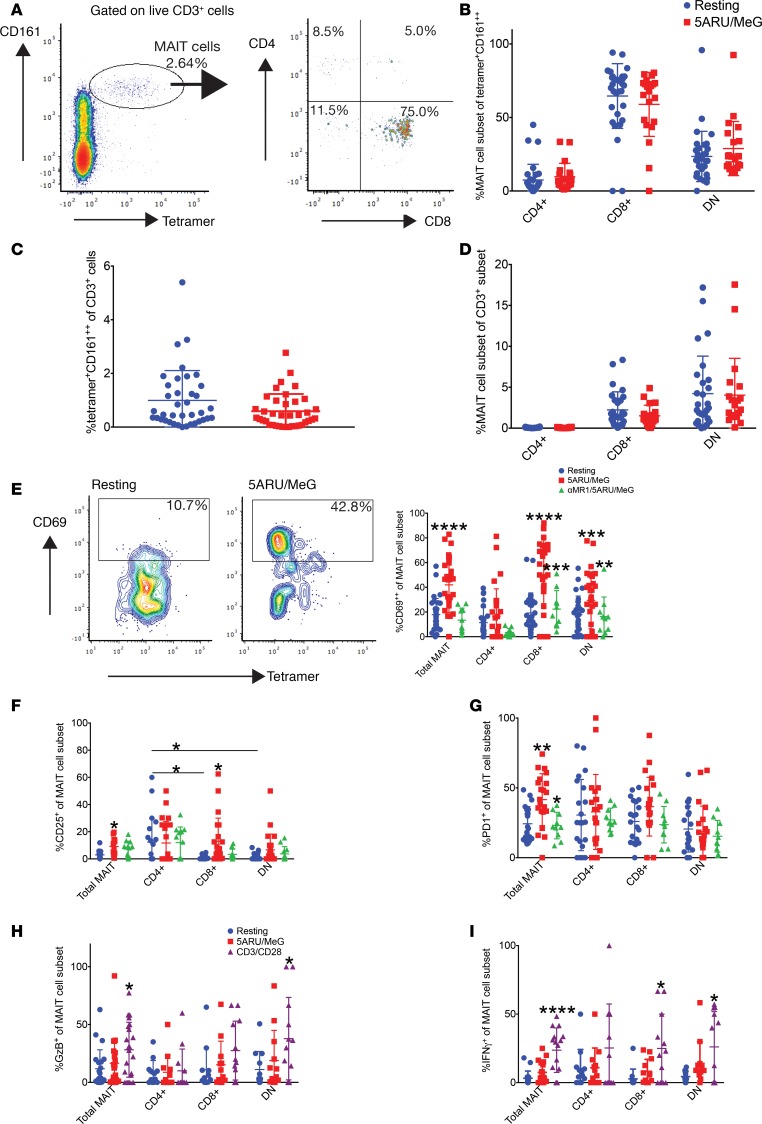
Figure 2. MAIT cell subsets demonstrate functional heterogeneity in healthy Haitian donors.
(A) Density plots demonstrating the gating strategy for MAIT cells in 1 healthy Haitian donor: left panel is gated on live CD3+ cells, and right panel is gated on live CD3+MR1-5-A-RU/MeG tetramer+CD161++. (B) Relative abundance of MAIT cell subsets among tetramer+CD161++ cells after 15 hours of rest (blue) or 5-A-RU/MeG activation (red) in 32 healthy Haitian donors. Same donors assessed in B–I. Same color coding applies to C and D. Data in B–D and F–I represent mean ± SD. (C) Abundance of MAIT cells among T cells. (D) Abundance of MAIT cells within their respective T cell subset. (E) Contour plots demonstrating the gating strategy for MAIT cell CD69 staining, a T cell activation marker, at rest or after 5-A-RU/MeG (left panels). Both plots are gated on live, CD3+, tetramer+CD161++. The right panel demonstrates CD69 staining in MAIT cell subsets at rest (blue), after 5-A-RU/MeG activation (red), or after treatment with neutralizing αMR1 antibody for 1 hour prior to 5-A-RU/MeG activation (green). Same color coding applies to F and G. (F) CD25 and (G) PD-1 staining in MAIT cell subsets. (H) GzB and (I) IFNγ measured by intracellular staining in the resting (blue), 5-A-RU/MeG (red), and anti-CD3/CD28 (purple) conditions in MAIT cell subsets. Groups were compared by 2-tailed unpaired t test with a significance level of P < 0.05. Asterisks above stimulated conditions indicate statistical significance compared with resting. Asterisks above the αMR1 condition indicate statistical significance compared with the 5-A-RU/MeG-stimulated condition. *P < 0.05, **P < 0.005, ***P < 0.0005, ****P < 0.0001; 5-A-RU, 5-amino-6-(D-ribitylamino)uracil; MeG, methylglyoxal; GzB, granzyme B.

