Figure 2.

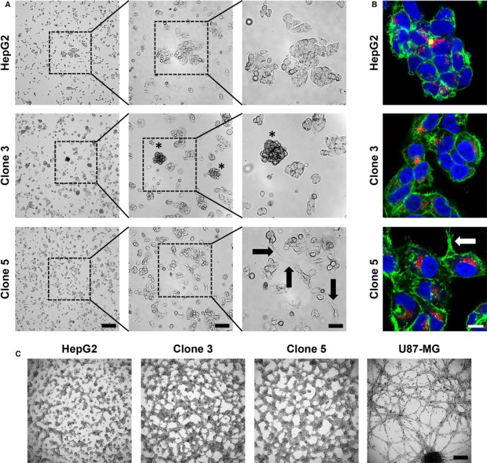
Cell morphology, growth pattern and tube formation ability of HepG2, clone 3 and clone 5 cells. A, Exemplary light microscopy images of HepG2, clone 3 and clone 5 cells cultured in 2D after 48 h of incubation (0 h—seeding of 1 × 10⁶ cells in a 10 cm culture dish). The experiment was carried out in triplicate. 4×—250 μm, 10×—100 μm, 20×—50 μm. Asterisks—spheroid‐like clusters; Arrows—elongated cell protrusions. B, Representative immunofluorescence staining images of HepG2, clone 3 and clone 5 cells. Green—F‐actin; red—AFP; blue—nuclei. Arrow—elongated cell protrusion. Scale—10 μm. C, Tube Formation assay to assess the ability of HepG2, clone 3 and clone 5 cells to form vasculogenic mimicry after growth on Matrigel for 24 h. Scale—250 μm. All images are representative for at least two independent experiments
