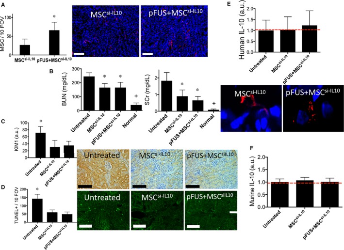
Figure 3.

IL‐10‐silenced mesenchymal stromal cells (MSCs) infused into wild‐type C3H mice following pFUS to kidneys do not increase IL‐10 expression or improve AKI outcomes compared to infusions of IL‐10‐silenced MSCs alone. C3H mice with AKI were given i v infusions of 106 IL‐10‐silenced human MSC (MSC si‐ IL 10) with or without pFUS (n = 6 mice for all experimental groups). (A) pFUS significantly increased MSC si‐ IL 10 homing to AKI kidneys (P < 0.05). Fluorescence IHC for human mitochondria is shown to the right (scale bar = 100 μm; MSC = red; nuclei = blue). (B) Renal function (BUN and SCr clearance) in C3H mice was significantly improved by MSC si‐ IL 10 alone (P < 0.05 compared to untreated controls), but additional significant reductions were not observed when mice were treated with pFUS+MSC si‐ IL 10 (P > 0.05 compared to MSC alone). (C) Renal KIM‐1 expression, D) TUNEL+ cells during AKI in C3H mice are significantly improved by infusion of MSC si‐ IL 10 alone (P < 0.05 compared to untreated controls), but not further reduced (P > 0.05 compared to MSC alone) with pFUS+MSC si‐ IL 10. Representative IHC shown at right (scale bar = 100 μm). (E) Human IL‐10 expression remained undetectable in kidneys from mice that received pFUS+MSC si‐ IL 10 or MSC si‐ IL 10 alone. For reference, kidneys from untreated C3H mice (AKI, but no MSC or pFUS) are shown with the red line to indicate assay background levels. IHC staining of human mitochondria (red) and human IL‐10 (green) revealed no increased IL‐10 expression in MSC si‐ IL 10 following infusion into mice with pFUS‐pretreated kidneys. F) Murine IL‐10 levels were unchanged in C3H mice with AKI that received MSC alone or pFUS+MSC compared to untreated AKI mice. Groups with identical symbols are statistically similar and are statistically different from groups with different symbols
