Figure 6.

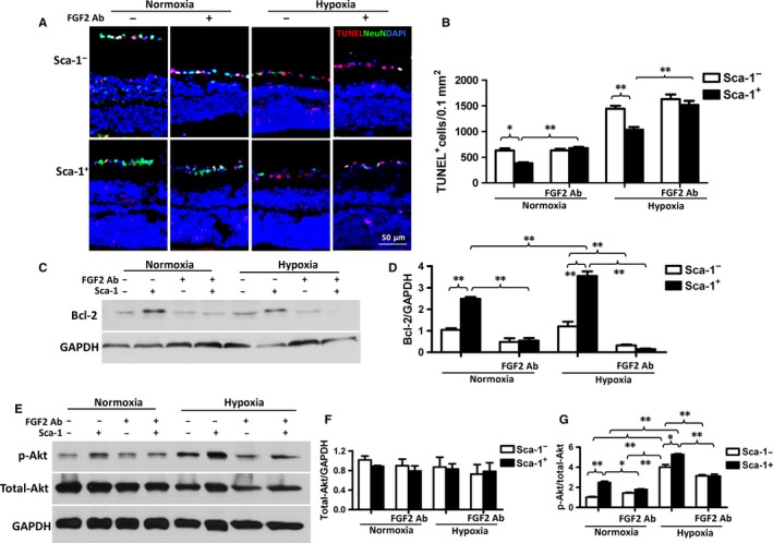
In vitro co‐culture of BM‐derived Sca‐1+ cells with retina explants decreased retinal cell apoptosis. Retina explants were co‐cultured with BM‐derived Sca‐1+ or Sca‐1− cells under normoxia and hypoxia conditions. (A) Representative images of apoptotic cells (Tunel+, red) and NeuN+ (green) neurons. Nuclei were stained with DAPI. (B) Co‐culture with BM Sca‐1+ cells protected the retina cells from hypoxia‐induced apoptosis. However, this protective effect was lost after application of an FGF2 (Fibroblast Growth Factor 2) neutralising antibody (Ab, n = 6/group). (C and D) Bcl‐2 protein expression was significantly greater in retina explants co‐cultured with BM Sca‐1+ than Sca‐1− cells under both normoxia and hypoxia conditions as assessed by Western blot. However, this effect was blocked by an FGF2 antibody (Ab). GAPDH was used as a loading control (n = 3/group). (E‐G) Total Akt and p‐Akt (phospho Ser473‐Akt) expression was assessed by Western blots. The ratio of p‐Akt/total Akt expression was significantly greater in retina explants co‐cultured with BM Sca‐1+ than Sca‐1− cells under both normoxia and hypoxia conditions. The ratio of p‐Akt/total Akt expression was significantly decreased after the application of an FGF2 Ab (n = 3/group). Data analysis used two‐way ANOVA followed by Bonferroni post‐hoc tests for multiple comparisons (B, D, F and G). Data shown are mean ± SEM. **P < 0.01, *P < 0.05
