
Figure 3.
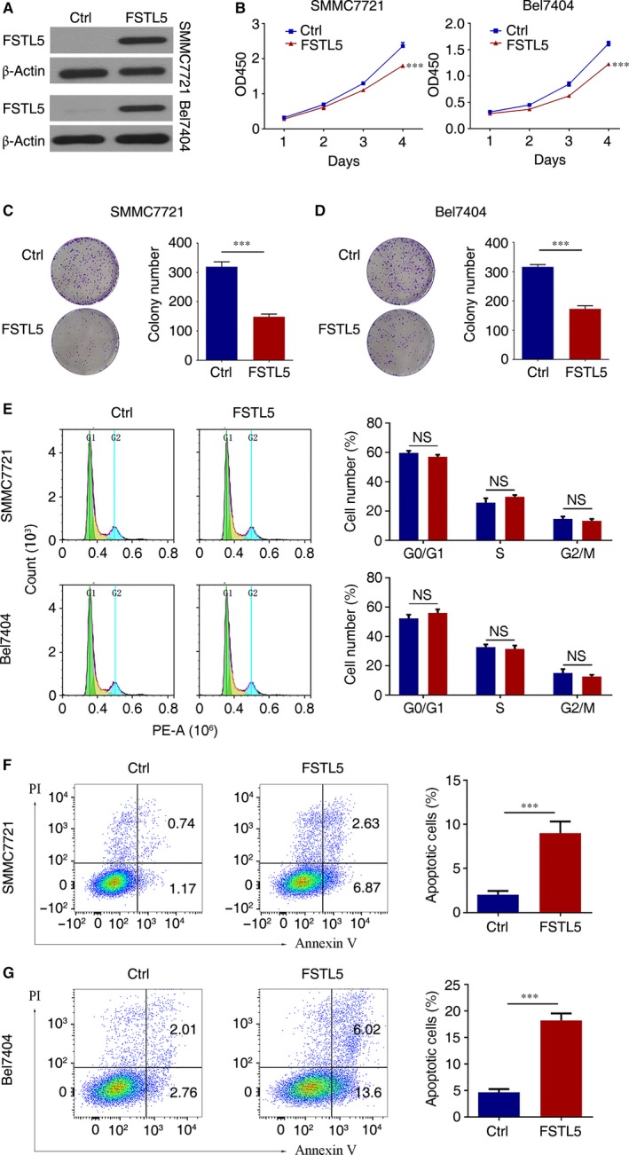
FSTL5 suppresses growth of HCC cell lines by inducing apoptosis in vitro. (A) Western blotting showing plasmid transfection efficiency. (B) CCK‐8 assay results showing the growth curve of SMMC7721/Bel7404 cells after transfection with pcDNA3.1(+)‐FSTL5 or pcDNA3.1(+) (n = 5, ***P < 0.001, Student's t test). (C and D) Colony formation assay of SMMC7721/Bel7404 cells after transfection with pcDNA3.1(+)‐FSTL5 or pcDNA3.1(+) (n = 3, ***P < 0.001, Student's t test). (E) Flow cytometry assay to determine cell cycle stages of SMMC7721/Bel7404 cells 72 hours after transfection with pcDNA3.1(+)‐FSTL5 or pcDNA3.1(+) (n = 3, NS, Student's t test). (F and G) Flow cytometry assay for apoptosis of SMMC7721/Bel7404 48 hours after transfection with pcDNA3.1(+)‐FSTL5 or pcDNA3.1(+) (n = 3, ***P < 0.001, Student's t test)