Figure 7.

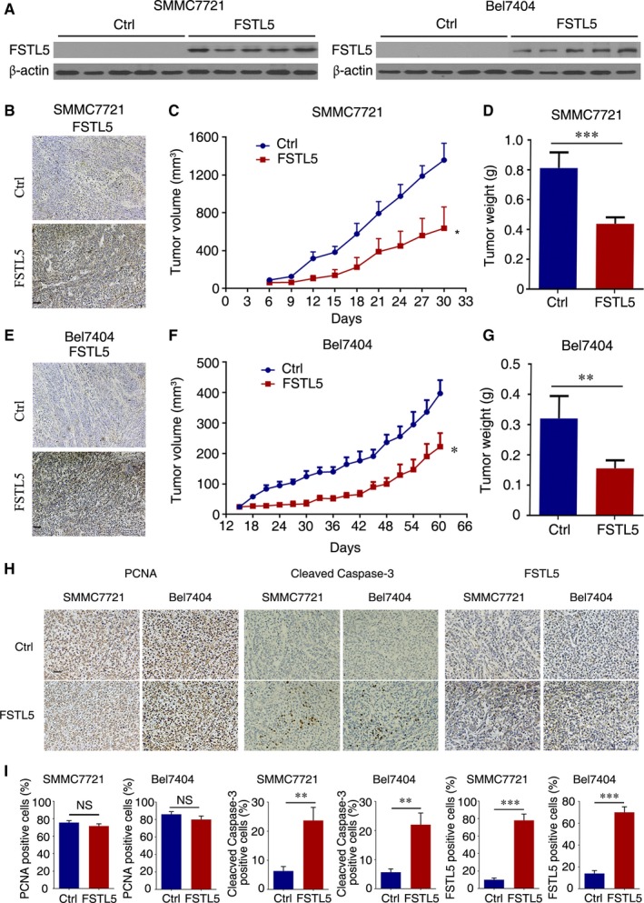
FSTL5 inhibits HCC tumor growth in vivo. (A) Western blotting showing FSTL5 expression in FSTL5 and control SMMC7721/Bel7404 xenografts. (B and E) IHC staining for FSTL5 in FSTL5‐expressing and control SMMC7721/Bel7404 xenografts (scale bar = 50 μm). (C and D) Tumor volume (n = 5, *P < 0.05, Student's t test), and end‐stage tumor weight (n = 5, ***P < 0.001, Student's t test), after injection of FSTL5‐expressing and control SMMC7721 cancer cells into nude mice. (F and G) Tumor volume (n = 5, *P < 0.05, Student's t test), and end‐stage tumor weight (n = 5, **P < 0.01, Student's t test), after injection of FSTL5‐expressing and control Bel7404 cancer cells into nude mice. (H and I) IHC staining for PCNA, Cleaved Caspase‐3 and FSTL5 in xenografts from nude mice (n = 3, **P < 0.01, ***P < 0.001, Student's t test, scale bar = 25 μm)
