Figure 3.

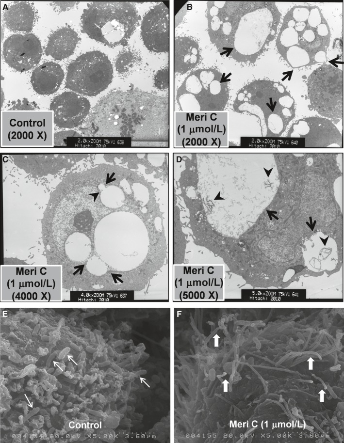
Transmission and scanning electron micrographs of meridianin C‐induced macropinosomes and macropinocytosis in YD‐10B cells. (A‐D) YD‐10B cells were treated with meridianin C (1 μM) or vehicle control (DMSO) for 8 h. (A) Vehicle (DMSO)‐treated cells, 2000 X. (B) Meridianin C‐treated cells, 2000 X. Arrows indicate the cytoplasmic vacuoles of different sizes. (C) Meridianin C‐treated cells, 4000 X. Arrowhead indicates the indented or distorted nucleus because of enlarged vacuole. Arrows indicate fusion between cytoplasmic vacuoles of different sizes. (D) Meridianin C‐treated cells, 5000 X. Arrows indicate macropinosomes containing membranous contents in different sizes, which are shown by arrowheads. (E, F) YD‐10B cells were treated with meridianin C (1 μM) or vehicle control (DMSO) for 2 h. (E) Vehicle control treated cells, which show many short and thick bacilli bound to the cell surface (thin arrows). (F) Meridianin C‐treated cells, which exhibit abundant long and thin membrane extensions that resemble lamellipodia on the cells surface (wide white arrows)
