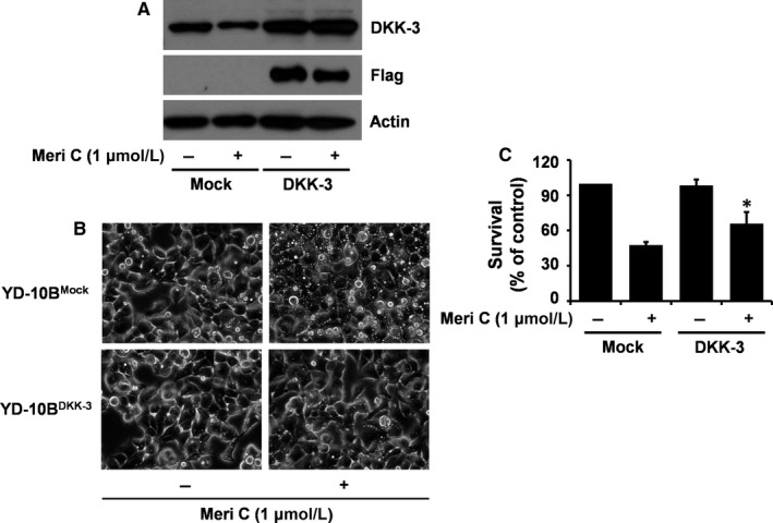
Figure 7.

Effects of DKK‐3 overexpression on meridianin C‐induced accumulation of vacuoles and decrease in survival of YD‐10B cells. (A) YD‐10B cells were transfected with pcDNA3.1‐DKK‐3‐Flag or mock vector using LipofectAMINE 2000 reagents. Stable clones were selected in the culture media containing 200 μg/ml G418 sulphate for several weeks. The selected clones (DKK‐3‐Flag or mock vector‐transfected YD‐10B cells) were seeded overnight. Cells were then treated with or without meridianin C (1 μM) for 20 min. Cell lysates were prepared and analysed by Western blotting for measurement of the expression levels of DKK‐3, Flag and β‐actin. (B) DKK‐3‐ or mock‐transfected YD‐10B cells were treated vehicle or meridianin C (1 μM) for 4 h. Images were obtained by phase contrast microscopy, 400 X. Each image is a representative of three independent experiments. (C) DKK‐3‐ or mock‐transfected YD‐10B cells were treated with or without meridianin C (1 μM) for 24 h, followed measurement of the number of cells survived by the trypan blue dye exclusion assay. Data are mean ± SEM of three independent experiments, each performed in triplicate. *P < 0.05 compared to the control at the indicated time
