
Figure 2. Direct activation of spinal TLR4 by KLA in vivo significantly increases hepoxilin production in lumbar spinal cord tissue and microglial cultures.
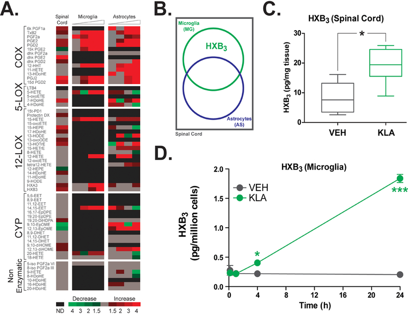
(A) Timecourse array of changes in levels of COX, nonenzymatic, LOX and CYP metabolites measured in lumbar spinal cord tissue at 2h and released from cultures of spinal microglia and astrocytes at baseline, 2h and 8h after treatment with VEH (IT saline or 0.1% DMSO, respectively) or KLA (1 μg/10 μl IT or 100 ng/ml, respectively). Fold differences are indicated on the heat map as follows: green, decreased; red, increased; gray, no change; black, not detected. (B) Metabolite changes following KLA treatment were analyzed using the following criteria (illustrated in diagram): not produced by COX (due to NSAID insensitivity), significantly (P<0.05) and robustly (≥ 2-fold) altered in both spinal cord tissue and cultured glial cells (microglia, astrocytes, or both). These criteria reveal a single lipid target (HXB3) that is increased in spinal cord (C) and microglia (D) following treatment with KLA. *P<0.05, ***P<0.001, n = 8–15 rats/group or n = 4 wells/treatment. VEH, vehicle; KLA, KDO2 Lipid A; COX, cyclooxygenase; LOX, lipoxygenase; CYP, cytochrome P450.