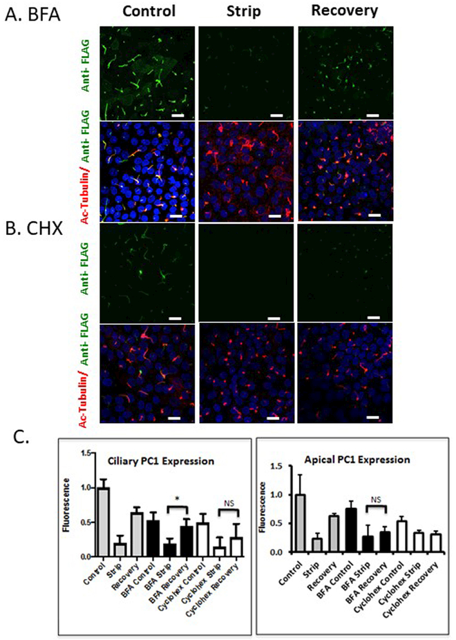
Figure 3: BFA treatment inhibits PC1 delivery to the apical plasma membrane but not to the cilium.
A and B LLC-PK1 cells stably expressing PC1 and PC2 were subjected to the alkaline stripping protocol to remove the Flag-tagged PC1 N-terminus, and then allowed to recover for 7 hours. Cells treated with BFA (A) or CHX (B) received drug treatment for 2 hours preceding strip, and during the 7 hours of recovery. Anti-Flag antibody staining is presented in a z-stack of confocal images counter-stained with anti-acetylated tubulin to identify the cilia. C Quantification of average pixel intensity of n=300 cilia (left panel) shows ciliary recovery in control conditions and cells treated with BFA. Significant apical recovery (right panel, * p<0.05) was detected under control conditions, but not in cells treated with BFA. No significant apical recovery was detected in CHX treated cells, but modest ciliary recovery persisted. All scale bars correspond to 25 μ. N=3

