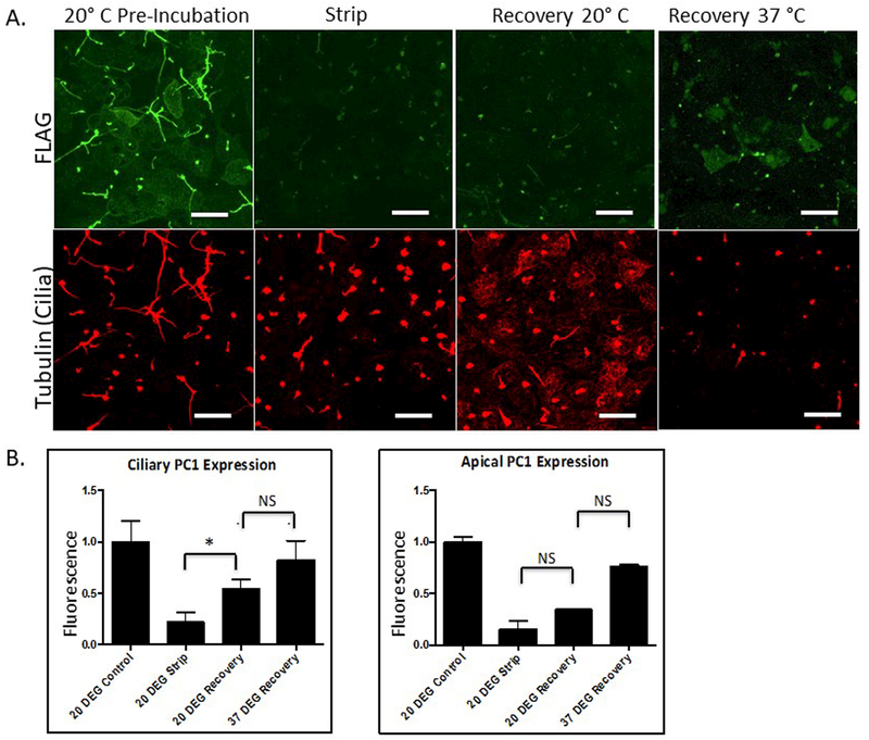
Figure 4: Incubating cells at 20oC substantially slows apical PC1 recovery but does not prevent ciliary recovery.
Strip-Recovery was performed on LLC-PK1 cells expressing PC1 and PC2. Cells were incubated at 20o C for 1 hour before incubation in the alkaline stripping buffer. Post strip, cells were incubated at either 20°C or 37 °C. A Surface expression of PC1 was detected under the different conditions by surface immunofluorescence utilizing an anti-Flag antibody to detect the extracellular Flag epitope on the N-terminus of PC1. Cilia were detected using an antibody directed against acetylated tubulin. B Quantitation of data from experiments following the design depicted in A. Error bars represent the SEM. (* p<0.013) All scale bars correspond to 25 μ. N=3

