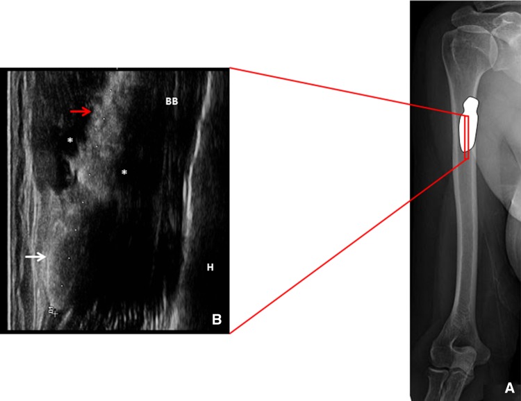
Fig. 1.
Position of the probe at the proximal third of the biceps brachii muscle in the long axis view (a). The corresponding ultrasound image shows the calcium deposit located over the superficial fascia of the biceps brachii muscle with a caudal crystalline portion elliptical in shape (white arrow), and a cranial hydrated portion, more irregular in shape, slipped between the fasciae (red arrow) with peri-calcific edema (*) (b)

