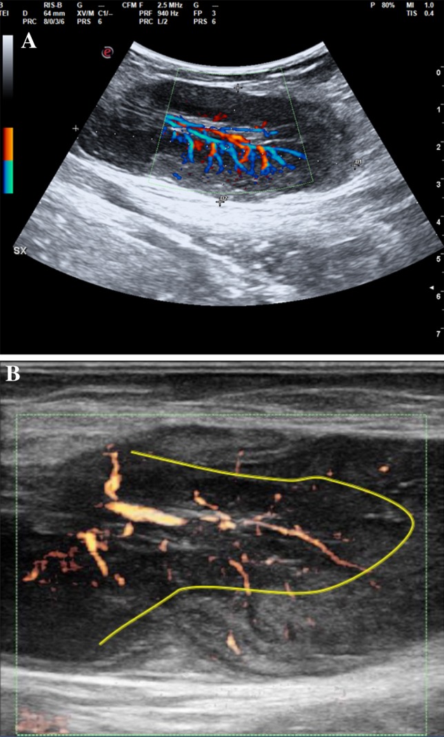
Fig. 1.

Pathological lymph node displaying hypertrophic hilar vessels; the perfusion stops its transition between the medullary and cortical areas, thus depriving most of the peripheral area of its physiological vascularization. a Color Doppler analysis; b power Doppler analysis
