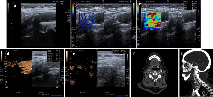
Fig. 1.
a At baseline US, a mainly calcified plaque was detected at right carotid bifurcation. b SWE quality control shows that the sampling was done correctly. c SWE confirms that it is a hard plaque (93.7 kPa). d, e CEUS in axial and longitudinal view allows a clear evaluation of the grade of stenosis; no significant focus of contrast enhancement is visible within the plaque; an ulceration is visible (better seen in longitudinal sonogram). f, g CTA in axial and sagittal (MIP) view; plaque is mixed, ulceration is confirmed; no significant contrast enhancement is detected

