Figure 9.
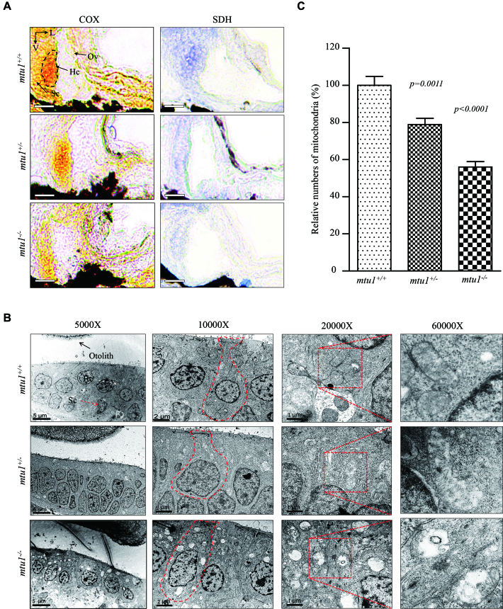
Mitochondrial defects in hair cells. (A) Assessment of mitochondrial function in hair cells by enzyme histochemistry (EHC) staining for SDH and COX in the frozen-sections of posterior macula of mtu1−/−, mtu1+/− and mtu1+/+ zebrafish at 5dpf. Loss of EHC signal is indicated by arrows (magnification X400). V, ventral; L, lateral; Ov, otic vesicle; Hc, hair cell; Sc, supporting cell. (B) Mitochondrial networks from hair cells of inner electron microscopy. Ultrathin sections were visualized with 5000×, 10 000×, 20 000× and 60 000× magnifications. (C) Quantification of mitochondrial numbers of hair cells from the mtu1−/−, mtu1+/− mutant and mtu1+/+ zebrafish. The calculations were based on 50 different hair cells of mtu1−/−, mtu1+/− mutant and mtu1+/+ zebrafish, respectively. Graph details and symbols are explained in the legend to Figure 3.

