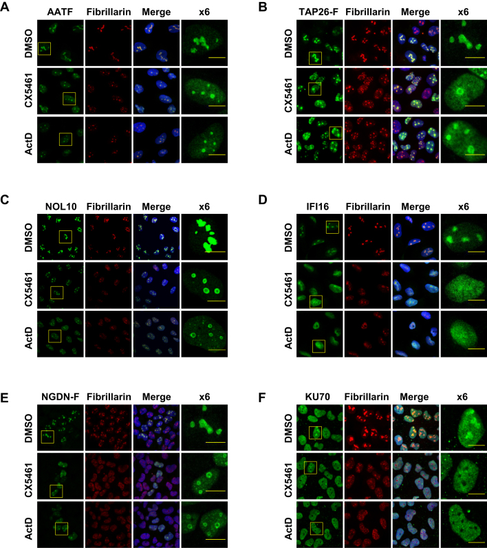
Figure 3.
Subcellular distribution of AATF, TAP26, NOL10, IFI16, NGDN and KU70 in control and ActD or CX5461-treated cells. Immuno-localisation of AATF (A), FLAG-tagged TAP26 (B), NOL10 (C), IFI16 (D), FLAG-tagged NGDN (E) and KU70 (F) in U-2 OS cells. Fibrillarin and Hoechst staining were used as nucleolar and nuclear markers respectively. The subcellular distribution of each protein was monitored in control cells and in cells treated with actinomycin D (ActD) or CX5461. Each of the candidate RNAPI-dependent RBPs is present in the nucleolus. The size bar represents 10 microns.

