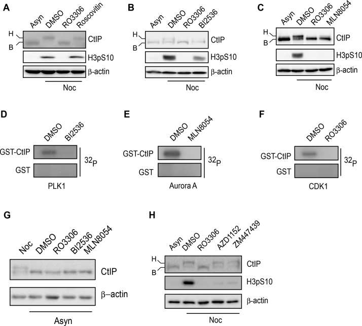
Figure 2.
CtIP is hyperphosphorylated by mitotic kinase. (A–C) U2OS cells were incubated with nocodazole or DMSO for 16 h and then treated with the indicated drugs for a further 1 h. After drug treatment, the cells were collected, lysed and subjected to western blotting using the indicated antibodies. (D–F) Purified GST-CtIP proteins from Sf9 insect cells (Supplementary Figure S3A) were incubated with [γ-32P] ATP in the presence or absence of the indicated kinases or inhibitors for an in vitro kinase assay. The radio-labeled CtIP was visualized following SDS-PAGE. (G) U2OS cells were incubated with nocodazole or the indicated drugs for 16 h. After drug treatment, the cells were collected, lysed and subjected to western blotting using the indicated antibodies. (H) The same experiment was performed as in (A–C). Asyn, asynchronized; B, basal CtIP; H, hyperphosphorylated CtIP; Noc, nocodazole .

